A METHOD FOR STUDYING THE MUTUAL ORIENTATION OF PLATE SURFACES MADE OF OPTICALLY TRANSPARENT MATERIALS DOI: https://doi.org/10.22184/1993-8578.2024.17.3-4.200.206
This paper describes a device for express measurement of plate tilt angles and angles between faces of plates transparent in the visible range in the visible radiation range. The range of values of angles, which can be measured by this setup, is calculated. Measurements and calculations of reflection coefficients of samples from silicon, sapphire, quartz and polymethyl methacrylate, the surfaces of which have different roughness, allowed us to formulate a restriction on the quality of the surface under study: the rms roughness should not exceed 50 nm.
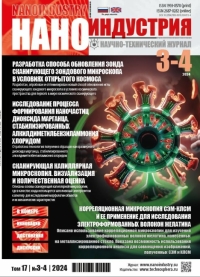
DEVELOPMENT OF A METHOD FOR UPDATING THE PROBE OF A SCANNING PROBE MICROSCOPE IN OPEN SPACE CONDITIONS | DOI: https://doi.org/10.22184/1993-8578.2024.17.3-4.166.175
A new method of updating the needle of a scanning probe microscope by sputtering different materials on it has been developed, tested and optimised. Experiments on sputtering different metals on the needle were performed, scans were taken with this needle in a scanning tunnelling microscope before and after sputtering metals on it, and the change in resolving power after sputtering metals as the main parameter of the microscope was evaluated. The new method of probe updating was developed for the world’s first space scanning tunnelling microscope, but it is also applicable for probe microscopes of other types, for example, for atomic force microscopes, as well as for various applications of probe microscopes in general, including those operating in vacuum chambers for various purposes.
FEMTOSCAN ONLINE: IMAGE PROCESSING AND FILTERING DOI: https://doi.org/10.22184/1993-8578.2024.17.3-4.178.183
The unique capabilities of the atomic force microscope (AFM), including super-high-resolution imaging, nanomanipulation, and the ability to operate under physiological conditions, have opened up exciting research opportunities in biology and biomedicine. AFM imaging has helped reveal the fine structure of bacterial cell walls at the nanoscale and how they are altered by antimicrobial treatment. This paper discusses the functions of FemtoScan Online software in relation to processing probe microscopy images, improving image quality reducing noise and increasing the informativeness of data.
SCANNING CAPILLARY MICROSCOPY. IMAGING AND QUANTIFYING DOI: https://doi.org/10.22184/1993-8578.2024.17.3-4.184.189
The study of the morphology of objects and their mechanical characteristics makes it possible to detect the unique properties of cells and associate these features with development under normal conditions or in the presence of pathologies. To measure the surface of a sample, scanning capillary microscopy (SCM) uses an electrolyte-filled capillary with a nano-sized hole at the tip as a probe. The main advantage of SCM is the non-contact visualization of the biological objects topography in the natural environment – scanning is carried out without forceful contact of the probe tip with the sample surface. Additionally, SCM can be used to determine electrical charges at the solid-liquid interface. In this article, we describe the basics of SCM, its capabilities for imaging cells, and measuring the biomechanical properties of living samples.
LABORATORY COMPLEX FOR OBTAINING COLLOIDAL PHOTONIC-CRYSTAL STRUCTURES. PART 1 DOI: https://doi.org/10.22184/1993-8578.2024.17.3-4.190.198
Colloidal photonic crystal structures are a promising material for nanoengineering. The goal of the work was to create a set of scalable equipment for the synthesis of monodisperse colloidal particles and the production of superlattices from them. The authors presented a description of the kit, the results of a study of the structures and formulated recommendations for the design of equipment and the implementation of technological processes.
SEM-CLSM CORRELATION MICROSCOPY AND ITS APPLICATION TO ELECTROSPUN GELATIN FIBERS DOI: https://doi.org/10.22184/1993-8578.2024.17.3-4.208.218
The most comprehensive information about microstructure of the sample can be obtained by combining different types of high-resolution microscopy. This combination turns out to be especially informative when measurements are carried out not only on the same image, but on the same area of the sample. This approach is called correlation microscopy. Typically, such experiments require careful preparation of the sample and transferring it between the two microscopes. The current work uses correlation microscopy which combines scanning electron microscopy (SEM) and confocal laser scanning microscopy (CLSM). Electrospun gelatin fibers deposited onto metallized glass are studied using these two methods. The possibility of using correlation analysis to combine images obtained by SEM and CLSM is demonstrated.
ORIGINALITY WITHOUT PRIVILEGE: COMPARATIVE ANALYSIS OF ELECTROFORMED AND STANDARD NITROCELLULOSE MEMBRANES IN MELATONIN IMMUNOASSAYS DOI: https://doi.org/10.22184/1993-8578.2024.17.3-4.220.229
Electrospinning enables manufacturing of polymer membranes composed of nanofibres. These membranes find application as filters, wound coatings, and tissue engineering scaffolds. They are also considered promising substrates for immunoassays. Despite the scientific community’s keen interest in the immunoassays based on electrospun membranes, a direct comparison with membranes formed through alternative technologies has not been conducted to date. In this study, we performed such a comparison and demonstrated that the detection of melatonin via enzyme-linked immunosorbent assay (ELISA) is virtually identical on the electrospun membranes and the conventional commercially available membranes.
STUDY OF THE FORMATION PROCESS OF MANGANESE DIOXIDE NANOPARTICLES STABILIZED BY ALKYLDIMETHYLBENZYLAMMONIUM CHLORIDE DOI: https://doi.org/10.22184/1993-8578.2024.17.3-4.230.239
Samples of nanosized manganese dioxide stabilized with alkyldimethylbenzylammonium chloride were obtained by chemical deposition. During optimization, it was revealed that for the synthesis of manganese dioxide nanoparticles with an average hydrodynamic radius of less than 1200 nm, the optimal synthesis parameters are: temperature from 20 to 35 °C, KMnO4 mass from 4 to 5 g and stabilizer concentration from 4 to 5%. Study of the samples using scanning electron microscopy showed that a sample of nano-sized manganese dioxide stabilized with alkyldimethylbenzylammonium chloride is formed by irregularly shaped aggregates ranging in size from 1 to 75 μm, which consist of nanoparticles with a diameter from 50 to 250 nm. The structure was studied using X-ray diffractometry and it was found that the resulting sample has a tetragonal crystal lattice with space group I4/m; presence of this phase is indicated by presence of low-intensity broadened peaks. As a result of analyzing the data obtained by modeling the interaction of the alkyldimethylbenzylammonium chloride molecule and manganese oxide through the nitrogen, it was established that the presented compounds are energetically favorable (∆E = 1299.571 kcal/mol), and interaction occurs through nitrogen. This compound has a chemical hardness value η ≥ 0.030 eV, which indicates its stability. As a result of the analysis of the IR-spectra of alkyldimethylbenzylammonium chloride and nano-sized manganese dioxide stabilized by alkyldimethylbenzylammonium chloride, it can be concluded that interaction between alkyldimethylbenzylammonium chloride and manganese dioxide occurs through nitrogen.

 rus
rus


